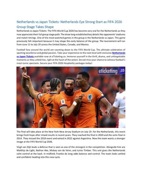
[ad_1]
Suggest an improvement

/* “function”==typeof InitializeEditor,callIfLoaded:function(o)return!(!gform.domLoaded,initializeOnLoaded:function(o)(document.addEventListener(“gform_main_scripts_loaded”,()=>gform.scriptsLoaded=!0,gform.callIfLoaded(o)),document.addEventListener(“gform/theme/scripts_loaded”,()=>gform.themeScriptsLoaded=!0,gform.callIfLoaded(o)),window.addEventListener(“DOMContentLoaded”,()=>gform.domLoaded=!0,gform.callIfLoaded(o))),hooks:action:,filter:,addAction:function(o,r,e,t)gform.addHook(“action”,o,r,e,t),addFilter:function(o,r,e,t)gform.addHook(“filter”,o,r,e,t),doAction:function(o)gform.doHook(“action”,o,arguments),applyFilters:function(o)return gform.doHook(“filter”,o,arguments),removeAction:function(o,r)gform.removeHook(“action”,o,r),removeFilter:function(o,r,e)gform.removeHook(“filter”,o,r,e),addHook:function(o,r,e,t,n)null==gform.hooks[o][r]&&(gform.hooks[o][r]=[]);var d=gform.hooks[o][r];null==n&&(n=r+”_”+d.length),gform.hooks[o][r].push(tag:n,callable:e,priority:t=null==t?10:t),doHook:function(r,o,e)var t;if(e=Array.prototype.slice.call(e,1),null!=gform.hooks[r][o]&&((o=gform.hooks[r][o]).sort(function(o,r)return o.priority-r.priority),o.forEach(function(o)”function”!=typeof(t=o.callable)&&(t=window[t]),”action”==r?t.apply(null,e):e[0]=t.apply(null,e))),”filter”==r)return e[0],removeHook:function(o,r,t,n)var e;null!=gform.hooks[o][r]&&(e=(e=gform.hooks[o][r]).filter(function(o,r,e)return!!(null!=n&&n!=o.tag),gform.hooks[o][r]=e)});
/* ]]> */

-
N/AFix spelling/grammar issueAdd or fix a linkAdd or fix an imageAdd more detailImprove the quality of the writingFix a factual error
-
You don’t need to tell us which article this feedback relates to, as we automatically capture that information for you.
-
This allows us to get in touch for more details if required.
-
Enter a five letter word in lowercase
#gform_wrapper_38 .gform_footer visibility: hidden; position: absolute; left: -100vw;
-
This field is for validation purposes and should be left unchanged.
/* = 0;if(!is_postback)return;var form_content = jQuery(this).contents().find(‘#gform_wrapper_38’);var is_confirmation = jQuery(this).contents().find(‘#gform_confirmation_wrapper_38’).length > 0;var is_redirect = contents.indexOf(‘gformRedirect(){‘) >= 0;var is_form = form_content.length > 0 && ! is_redirect && ! is_confirmation;var mt = parseInt(jQuery(‘html’).css(‘margin-top’), 10) + parseInt(jQuery(‘body’).css(‘margin-top’), 10) + 100;if(is_form)jQuery(‘#gform_wrapper_38’).html(form_content.html());if(form_content.hasClass(‘gform_validation_error’))jQuery(‘#gform_wrapper_38’).addClass(‘gform_validation_error’); else jQuery(‘#gform_wrapper_38’).removeClass(‘gform_validation_error’);setTimeout( function() /* delay the scroll by 50 milliseconds to fix a bug in chrome */ jQuery(document).scrollTop(jQuery(‘#gform_wrapper_38’).offset().top – mt); , 50 );if(window[‘gformInitDatepicker’]) gformInitDatepicker();if(window[‘gformInitPriceFields’]) gformInitPriceFields();var current_page = jQuery(‘#gform_source_page_number_38’).val();gformInitSpinner( 38, ‘https://geekymedics.com/wp-content/plugins/gravityforms/images/spinner.svg’, true );jQuery(document).trigger(‘gform_page_loaded’, [38, current_page]);window[‘gf_submitting_38’] = false;else if(!is_redirect)var confirmation_content = jQuery(this).contents().find(‘.GF_AJAX_POSTBACK’).html();if(!confirmation_content)confirmation_content = contents;jQuery(‘#gform_wrapper_38’).replaceWith(confirmation_content);jQuery(document).scrollTop(jQuery(‘#gf_38’).offset().top – mt);jQuery(document).trigger(‘gform_confirmation_loaded’, [38]);window[‘gf_submitting_38’] = false;wp.a11y.speak(jQuery(‘#gform_confirmation_message_38’).text());elsejQuery(‘#gform_38’).append(contents);if(window[‘gformRedirect’]) gformRedirect();jQuery(document).trigger(“gform_pre_post_render”, [ formId: “38”, currentPage: “current_page”, abort: function() this.preventDefault(); ]); if (event && event.defaultPrevented) return; const gformWrapperDiv = document.getElementById( “gform_wrapper_38” ); if ( gformWrapperDiv ) const visibilitySpan = document.createElement( “span” ); visibilitySpan.id = “gform_visibility_test_38”; gformWrapperDiv.insertAdjacentElement( “afterend”, visibilitySpan ); const visibilityTestDiv = document.getElementById( “gform_visibility_test_38” ); let postRenderFired = false; function triggerPostRender() if ( postRenderFired ) return; postRenderFired = true; gform.core.triggerPostRenderEvents( 38, current_page ); if ( visibilityTestDiv ) visibilityTestDiv.parentNode.removeChild( visibilityTestDiv ); function debounce( func, wait, immediate ) var timeout; return function() var context = this, args = arguments; var later = function() timeout = null; if ( !immediate ) func.apply( context, args ); ; var callNow = immediate && !timeout; clearTimeout( timeout ); timeout = setTimeout( later, wait ); if ( callNow ) func.apply( context, args ); ; const debouncedTriggerPostRender = debounce( function() triggerPostRender(); , 200 ); if ( visibilityTestDiv && visibilityTestDiv.offsetParent === null ) const observer = new MutationObserver( ( mutations ) => mutations.forEach( ( mutation ) => if ( mutation.type === ‘attributes’ && visibilityTestDiv.offsetParent !== null ) debouncedTriggerPostRender(); observer.disconnect(); ); ); observer.observe( document.body, attributes: true, childList: false, subtree: true, attributeFilter: [ ‘style’, ‘class’ ], ); else triggerPostRender(); } );} );
/* ]]> */

Introduction
The elbow joint is a complex hinge-type synovial joint made up of articulations between the distal humerus and the proximal ulna and radius.1 This allows for significant movement and function of the forearm, including flexion and extension.1 The radioulnar joint facilitates supination and pronation of the hand.2

Bony structure
Distal humerus
The capitellum is a rounded bony prominence located at the lateral aspect of the distal humeral condyle.3 It covers the anterior and inferior surfaces of the lateral condyle. The capitellum articulates with the proximal radial head to form the radiohumeral joint.
The trochlear occupies a large portion of the medial part of the distal humerus.3 It is shaped like a pulley and covers the anterior, posterior, and inferior surfaces of the medial condyle. It articulates with the trochlear notch of the ulna to form the ulnohumeral joint. A small groove separates the trochlear from the capitellum.
The medial epicondyle is a prominent, non-articular bony projection of the medial condyle.4 It is larger than the lateral epicondyle and easily palpable. It serves as the attachment for the ulnar collateral ligament of the elbow joint and the superficial muscles of the anterior forearm. The ulnar nerve runs down a groove posterior to the medial epicondyle to innervate the muscles of the forearms.
The lateral epicondyle of the humerus is a non-articular bony eminence of the lateral condyle.4 It serves as the attachment for the radial collateral ligament and muscles of the posterior forearm.

Fossa
The three fossae at the distal humerus are:
- Radial fossa: superior to the capitellum. In full flexion, the radial fossa receives the head of the radius.
- Coronoid fossa: superior to the trochlear. In full flexion, the coronoid fossa receives the coronoid process of the ulna.
- Olecranon fossa: superior to the posterior surface of the trochlear. In full extension, the olecranon fossa receives the olecranon process of the ulna.4
Proximal radius
The head of the radius is cylindrical and is covered with hyaline cartilage.1 The upper surface of the articular circumference is concave and articulates with the capitellum. The medial aspect articulates with the radial notch of the ulna to form the radioulnar joint.
The radial tuberosity is an oval bony prominence on the medial aspect of the proximal part of the radius, distal to the neck of the radius.2 The tendon of the biceps brachii attaches to the posterior part of the radial tuberosity, and a bursa lies against the anterior surface.

Proximal ulna
The ulna is a long bone of the forearm located medially to the radius in the anatomical position.1 It has a large proximal end and tapers down to a smaller distal end. The ulnar articulates with the humerus and the radius proximally to stabilise the elbow joint.
The olecranon is a prominent and proximal extension of the ulnar shaft posteriorly.4 The top of the olecranon is shaped like a beak and lodges into the olecranon fossa of the humerus in elbow extension. This prevents hyperextension of the elbow joint.
The coronoid process is an anterior and forward projection at the proximal end of the ulnar shaft. It is lodged into the coronoid fossa of the distal humerus during flexion of the elbow. Medial to the lip of the coronoid process is the sublime tubercle, where the medial collateral ligament of the elbow attaches. The ulnar tuberosity is just distal to the coronoid process where the brachialis muscle inserts.
The trochlear notch is a concave and saddle-shaped surface located between the olecranon and the coronoid process. It is covered by hyaline cartilage and articulates with the trochlear of the humerus, allowing elbow flexion and extension.
The radial notch is a small concave surface located lateral to the coronoid process. It is covered by hyaline cartilage and articulates with the radial head. The annular ligament attaches around the anterior and posterior surfaces of the radial notch.
Ossification centres
The elbow has six ossification centres, which develop at different ages in a relatively reproducible manner.4 They can be easily mistaken for fractures, therefore, it is important to recognise and differentiate ossification centres from fractures in the paediatric population.
The order and age at which the ossification centres appear can be remembered by CRITOE:
- Capitellum: 2-24 months
- Radial head: 3-6 years
- Internal (medial) epicondyle: 4-7 years
- Trochlea: 8-10 years
- Olecranon: 8-10 years
- External (lateral) epicondyle: 10-13 years3
For example, in a 5-year-old child with an elbow injury, if the trochlea is visible on a radiograph but the medial epicondyle is not, the ossification centre of the medial epicondyle has most likely been avulsed and displaced.

Clinical relevance: Monteggia fracture and Galeazzi fracture
These are common fractures that occur from a fall on an outstretched hand (FOOSH). Due to the strong connection between the shaft radius and the ulna by the interosseous membrane, a fracture of one bone can result in the dislocation of the interconnected joint.
Management is open reduction and internal fixation of the radius and ulna.
Monteggia fracture: proximal ulnar fracture and proximal radial head dislocation.3

Galeazzi fracture: distal radial fracture and distal radioulnar joint dislocation.3

Articular surfaces
The articular surfaces are lined with hyaline cartilage.1 They are encapsulated in a fibrous joint capsule lined by synovial tissue.
There are three articulations of the elbow joint: the radiohumeral, ulnohumeral and radioulnar.1
Radiohumeral
- Articular surfaces: radial head and capitellum of the humerus
- Joint: hinge-type synovial joint
- Movement: flexion and extension at the elbow
Ulnohumeral
- Articular surfaces: trochlea of the humerus and the trochlear notch of the ulna
- Joint: hinge-type synovial joint
- Movement: flexion and extension at the elbow
Radioulnar
- Articular surfaces: radial head with the radial notch of the ulna
- Joint: pivot-type synovial joint
- Movement: pronation and supination of the hand
Ligaments
A collection of strong ligaments stabilise the elbow joint.3
Collateral ligaments
The radial collateral ligament (lateral) extends from the lateral epicondyle of the humerus and blends with the annular ligament.
The ulnar collateral ligament (medial) extends from the medial epicondyle of the humerus to the coronoid process of the ulna. It has a triangular shape and consists of three parts: anterior, posterior and inferior bands.
Annular ligament
The annular ligament encircles the head of the radius to hold it in place against the proximal ulna and to stabilise the proximal radioulnar joint.
Quadrate ligament
The quadrate ligament is located inferior to the annular ligament. It attaches to the distal margin of the radial notch and to the neck of the radius to stabilise the proximal radioulnar joint.

Bursae
Bursae are synovial fluid-filled sacs. They act as shock absorbers, providing a cushion between the bones and surrounding soft tissues.
There are three olecranon bursae:1
- Subcutaneous olecranon bursa: in the subcutaneous connective tissue over the olecranon
- Subtendinous olecranon bursa: between the olecranon and triceps tendon
- Intratendinous olecranon bursa: in the tendon of triceps brachii1
The bicipitoradial bursa or biceps bursa reduces friction between the distal biceps brachii tendon and the tuberosity of the radius

Blood supply and innervation
Blood supply
The arteries supplying the elbow joint are derived from the peri-articular anastomosis around the elbow joint.2 They originate from the brachial, profunda brachii, radial, and ulnar arteries, as well as their collateral and recurrent branches.

Innervation
The elbow joint is innervated by the musculocutaneous, median, radial, and ulnar nerves.2
Muscles
As a hinge joint, the elbow joint is responsible for flexion and extension of the forearm.3 Pronation and supination do not occur at the elbow joint, as these movements are afforded at the radioulnar joint.
Anterior compartment
Flexion at the elbow joint is produced by the two muscles of the anterior compartment of the arm, biceps brachii and brachialis.3 They are both innervated by the musculocutaneous nerve.
The biceps brachii is a powerful flexor of the elbow joint when the forearm is supinated and the elbow is flexed close to 90°. The two heads originate from the supraglenoid tubercle (long head) and the coracoid process (short head) of the scapula. They join together to form the muscle belly, and the biceps tendon inserts distal to the elbow joint at the radial tuberosity. The biceps brachii does not have any attachment to the humerus.
The brachialis lies posterior to the biceps brachii and is the main flexor of the forearm.
Posterior compartment
The triceps brachii and aconeus are the muscles of the posterior compartment of the arm and provide extension at the forearm, and are both innervated by the radial nerve.3
The triceps brachii is the main extensor of the forearm. It has three heads, as the name indicates. The long head originates from the infraglenoid tubercle of the scapula, the lateral head originates superolateral to the radial groove, and the medial head originates inferomedial to the radial groove. Together, they converge to the triceps tendon and insert onto the olecranon. The anconeus assists the triceps to extend the forearm and stabilise the elbow joint.

Clinical relevance: epicondylitis
Epicondylitis is inflammation of the soft tissues at the epicondyles of the humerus. It is commonly caused by overuse of the muscles of the forearm. Pain is often localised to either the medial or lateral epicondyle region.
Lateral epicondylitis (tennis elbow) results in lateral epicondylar pain where the common extensor muscles of the forearm attach.3
Medial epicondylitis (golfer’s elbow) results in medial epicondylar pain where the common flexor muscles of the forearm attach.3
Key points
- The elbow is a complex hinge-type synovial joint that plays a crucial role in the movement of the forearm and wrist
- Understanding the elbow ossification centres is crucial for distinguishing between normal developmental variations and fractures in paediatrics
- The elbow is formed by the three articulation surfaces between the distal humerus, the proximal ulna and the proximal radius
- The three pain ligaments stabilising the joint are the collateral, annular and quadrate ligaments
- The bursae provide a cushion between the bones and the surrounding soft tissue
- The elbow joint is responsible for flexion and extension of the forearm, with pronation and supination performed at the radioulnar joint
Reviewer
Orthopaedic Registrar
Editor
Dr Jamie Scriven
References
- Drake RL, Vogl WA, Mitchell AWM. Gray’s Anatomy for Students. Elsevier. 4th ed. 2019.
- OpenStax. Anatomy and Physiology. 2e. 2022. Available from: [LINK].
- Dalley AF, Agur AMR. Moore’s Clinically Oriented Anatomy. Wolters Kluwer Health. 9th ed. 2022.
- McMinn RMH. Last’s Anatomy. Churchill Livingstone Australia. 9th ed. 2019.
Image references
- Figure 1. OpenStax College. Figure 9.8 Synovial Joints. License: [CC BY 4.0].
- Figure 2. Andrew Murphy. Elbow anatomy (illustration). Radiopaedia. License: [CC BY-NC-SA 3.0].
- Figure 3. OpenStax College. Figure 8.6 Ulna and Radius. License: [CC BY 4.0].
- Figure 4. Andrew Dixon. Elbow radiograph – age eleven. Radiopaedia. License: [CC BY-NC-SA 3.0].
- Figure 5. Hellerhoff. Monteggia fracture. License: [CC BY-SA 3.0].
- Figure 6. Hellerhoff. Galeazzi fracture. License: [CC BY-SA 3.0].
- Figure 7. OpenStax College. Elbow joint. Adapted by Geeky Medics. License: [CC BY 3.0].
- Figure 8. OpenStax College. Elbow joint. Adapted by Geeky Medics. License: [CC BY 3.0].
- Figure 9. OpenStax College. Figure 20.31 Major Arteries Serving the Thorax and Upper Limb. License: [CC BY 4.0].
- Figure 10. OpenStax College. Figure 11.25 Muscles That Move the Forearm. Adapted by Geeky Medics. License: [CC BY 4.0].
[ad_2]
Source link
Discover more from Bibliobazar Digi Books
Subscribe to get the latest posts sent to your email.