[ad_1]
Suggest an improvement

/* “function”==typeof InitializeEditor,callIfLoaded:function(o)!gform.themeScriptsLoaded&&!gform.isFormEditor(),initializeOnLoaded:function(o),hooks:action:,filter:,addAction:function(o,r,e,t)gform.addHook(“action”,o,r,e,t),addFilter:function(o,r,e,t)gform.addHook(“filter”,o,r,e,t),doAction:function(o)gform.doHook(“action”,o,arguments),applyFilters:function(o)return gform.doHook(“filter”,o,arguments),removeAction:function(o,r)gform.removeHook(“action”,o,r),removeFilter:function(o,r,e)gform.removeHook(“filter”,o,r,e),addHook:function(o,r,e,t,n)null==gform.hooks[o][r]&&(gform.hooks[o][r]=[]);var d=gform.hooks[o][r];null==n&&(n=r+”_”+d.length),gform.hooks[o][r].push(tag:n,callable:e,priority:t=null==t?10:t),doHook:function(r,o,e)var t;if(e=Array.prototype.slice.call(e,1),null!=gform.hooks[r][o]&&((o=gform.hooks[r][o]).sort(function(o,r)return o.priority-r.priority),o.forEach(function(o)”function”!=typeof(t=o.callable)&&(t=window[t]),”action”==r?t.apply(null,e):e[0]=t.apply(null,e))),”filter”==r)return e[0],removeHook:function(o,r,t,n)var e;null!=gform.hooks[o][r]&&(e=(e=gform.hooks[o][r]).filter(function(o,r,e)return!!(null!=n&&n!=o.tag),gform.hooks[o][r]=e)});
/* ]]> */

-
N/AFix spelling/grammar issueAdd or fix a linkAdd or fix an imageAdd more detailImprove the quality of the writingFix a factual error
-
You don’t need to tell us which article this feedback relates to, as we automatically capture that information for you.
-
This allows us to get in touch for more details if required.
-
Enter a five letter word in lowercase
#gform_wrapper_38 .gform_footer visibility: hidden; position: absolute; left: -100vw;
-
This field is for validation purposes and should be left unchanged.
/* = 0;if(!is_postback)return;var form_content = jQuery(this).contents().find(‘#gform_wrapper_38’);var is_confirmation = jQuery(this).contents().find(‘#gform_confirmation_wrapper_38’).length > 0;var is_redirect = contents.indexOf(‘gformRedirect(){‘) >= 0;var is_form = form_content.length > 0 && ! is_redirect && ! is_confirmation;var mt = parseInt(jQuery(‘html’).css(‘margin-top’), 10) + parseInt(jQuery(‘body’).css(‘margin-top’), 10) + 100;if(is_form)jQuery(‘#gform_wrapper_38’).html(form_content.html());if(form_content.hasClass(‘gform_validation_error’))jQuery(‘#gform_wrapper_38’).addClass(‘gform_validation_error’); else jQuery(‘#gform_wrapper_38’).removeClass(‘gform_validation_error’);setTimeout( function() /* delay the scroll by 50 milliseconds to fix a bug in chrome */ jQuery(document).scrollTop(jQuery(‘#gform_wrapper_38’).offset().top – mt); , 50 );if(window[‘gformInitDatepicker’]) gformInitDatepicker();if(window[‘gformInitPriceFields’]) gformInitPriceFields();var current_page = jQuery(‘#gform_source_page_number_38’).val();gformInitSpinner( 38, ‘https://geekymedics.com/wp-content/plugins/gravityforms/images/spinner.svg’, true );jQuery(document).trigger(‘gform_page_loaded’, [38, current_page]);window[‘gf_submitting_38’] = false;else if(!is_redirect)var confirmation_content = jQuery(this).contents().find(‘.GF_AJAX_POSTBACK’).html();if(!confirmation_content)confirmation_content = contents;jQuery(‘#gform_wrapper_38’).replaceWith(confirmation_content);jQuery(document).scrollTop(jQuery(‘#gf_38’).offset().top – mt);jQuery(document).trigger(‘gform_confirmation_loaded’, [38]);window[‘gf_submitting_38’] = false;wp.a11y.speak(jQuery(‘#gform_confirmation_message_38’).text());elsejQuery(‘#gform_38’).append(contents);if(window[‘gformRedirect’]) gformRedirect();jQuery(document).trigger(“gform_pre_post_render”, [ formId: “38”, currentPage: “current_page”, abort: function() this.preventDefault(); ]); if (event && event.defaultPrevented) return; const gformWrapperDiv = document.getElementById( “gform_wrapper_38” ); if ( gformWrapperDiv ) const visibilitySpan = document.createElement( “span” ); visibilitySpan.id = “gform_visibility_test_38”; gformWrapperDiv.insertAdjacentElement( “afterend”, visibilitySpan ); const visibilityTestDiv = document.getElementById( “gform_visibility_test_38” ); let postRenderFired = false; function triggerPostRender() if ( postRenderFired ) return; postRenderFired = true; gform.core.triggerPostRenderEvents( 38, current_page ); if ( visibilityTestDiv ) visibilityTestDiv.parentNode.removeChild( visibilityTestDiv ); function debounce( func, wait, immediate ) var timeout; return function() var context = this, args = arguments; var later = function() timeout = null; if ( !immediate ) func.apply( context, args ); ; var callNow = immediate && !timeout; clearTimeout( timeout ); timeout = setTimeout( later, wait ); if ( callNow ) func.apply( context, args ); ; const debouncedTriggerPostRender = debounce( function() triggerPostRender(); , 200 ); if ( visibilityTestDiv && visibilityTestDiv.offsetParent === null ) const observer = new MutationObserver( ( mutations ) => mutations.forEach( ( mutation ) => if ( mutation.type === ‘attributes’ && visibilityTestDiv.offsetParent !== null ) debouncedTriggerPostRender(); observer.disconnect(); ); ); observer.observe( document.body, attributes: true, childList: false, subtree: true, attributeFilter: [ ‘style’, ‘class’ ], ); else triggerPostRender(); } );} );
/* ]]> */

Introduction
Interpreting musculoskeletal (MSK) X-rays can be challenging and is a skill commonly assessed in OSCEs. A systematic approach to interpreting MSK X-rays ensures that pathology is not missed.
Specific MSK X-ray findings and abnormalities commonly appear in OSCEs. You must be able to recognise these findings and discuss the management of these conditions.
These MSK X-rays may appear as part of data interpretation stations, ABCDE/simulation stations and communication stations.
Neck of femur fracture
A neck of femur fracture is a common fracture in elderly patients that has significant mortality. Patients typically present following a fall with a shortened and externally rotated leg, pain in the affected hip, and an inability to perform a straight-leg raise on the affected side.
Depending on the fracture location, they can be broadly classified into intracapsular and extracapsular, with intracapsular fractures carrying an increased risk of avascular necrosis of the femoral head.
Features of a neck of femur fracture on a hip X-ray may include:
- Interruption of Shenton’s line: loss of contour between the normally continuous line from the medial edge of the femoral neck and the inferior edge of the superior pubic ramus
- Displaced bony fragments
- Asymmetry when comparing the two femoral heads
- Flexed and externally rotated femur due to the unopposed action of iliopsoas, resulting in the lesser trochanter being more prominent
- A smudge-like appearance in impacted fractures


Management of neck of femur fractures
Initial management involves analgesia (frequently a fascia iliaca compartment block) and optimisation for surgery, which may include IV fluid resuscitation, blood transfusion, correcting electrolyte abnormalities, and reversing anticoagulation.
Surgical management depends on the location of the fracture, the patient’s functional baseline and past medical history:
- Intracapsular fractures are typically managed with a total hip replacement (replacing the femoral head and the acetabulum) or a hemi-arthroplasty (replacing only the femoral head)
- Extracapsular fractures are typically managed with fixation such as a dynamic hip screw (DHS) or intramedullary nail
Osteoarthritis
Osteoarthritis (OA) is a progressive, degenerative joint disorder. The most commonly affected joints are the knees, followed by the hips and hands. Typical symptoms include joint pain, stiffness that is typically worse after activity, reduced range of movement in affected joints, joint tenderness and crepitus on movement.
The cardinal features of OA on X-ray can be remembered with the acronym LOSS:
- Loss of joint space
- Osteophytes
- Subchondral sclerosis
- Subchondral cysts


Management of osteoarthritis
Initial management is conservative, including education and advice, physiotherapy, exercise, and weight loss (for those who are overweight). Medical management involves step-wise escalation of analgesia, with non-steroidal anti-inflammatory drugs (NSAIDs) preferred, if not contraindicated. Intrarticular steroid injections can also be beneficial for some.
Definitive management typically involves surgery, with most joints being treated through joint replacement (arthroplasty) or fusion (arthrodesis).
Colles’ fracture
A Colles’ fracture is the most common fracture of the distal radius. It is an extra-articular fracture with dorsal displacement of the distal fragment, causing a dinner fork deformity, and typically occurs following a fall on an outstretched hand (FOOSH).
Features of a Colles’ fracture on a wrist X-ray may include:
- An extra-articular fracture line visible through the distal radius
- A fracture line proximal to the radioulnar joint
- Impaction of the fracture and associated radial shortening
- Dorsal angulation of the distal fragment (seen as a diner fork deformity clinically if it is severe enough)
- Associated avulsion of the ulnar styloid process or carpal bone fractures


Management of Colles’ fracture
Management depends on the severity, the patient’s functional baseline, and co-morbidities.
The majority can be managed with manipulation and immobilisation in a below-elbow backslab for 4-6 weeks. Those with significant deformity or very complex fractures (e.g. comminuted fractures) may require open reduction and internal fixation (ORIF) with a plate and screws.
Once bone healing has occurred and plaster casts are removed, patients require physiotherapy to help regain function.
If this fracture occurs following a low-trauma injury, it would be considered a fragility fracture, and so assessment for possible osteoporosis should take place.
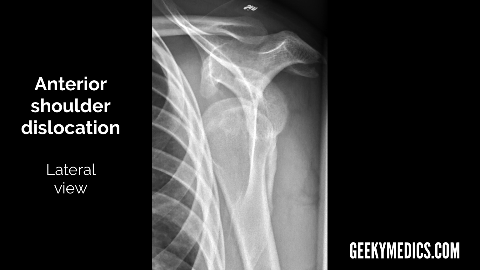
Shoulder dislocation
Shoulder dislocation occurs when there is complete dissociation of the humeral head from the glenoid fossa. The vast majority (95%) are anterior dislocations. Patients typically present with a painful shoulder with reduced range of movement, and the humeral head may be prominent anteriorly.
Imaging should always include an AP view and a scapular Y view.
Features of an anterior shoulder dislocation on shoulder X-ray may include:
- The humeral head lying medial and inferior to the glenoid fossa on an AP view
- The humeral head lying anterior and inferior to the glenoid fossa on a Y view
- The humeral head lying inferior to the coracoid process (typically most obvious in the lateral view)
- Associated fractures, such as Bankart lesions (damage to the glenoid labrum) or Hill-Sachs lesions (compression fractures of the posterolateral humeral head)


Management of shoulder dislocation
Management aims to reduce and relocate the joint. This can be carried out with local anaesthetic, conscious sedation or general anaesthesia depending on the setting and the patient. Conscious sedation should always be carried out in resus or theatre with monitoring and airway-trained staff.
A neurovascular examination should be performed before and after manipulation, and repeat imaging obtained after manipulation. The arm should be kept in a sling for two weeks, and follow-up with orthopaedics and physiotherapy should be organised.
Recurrent dislocations are common, and patients may require surgical intervention.
Ankle fracture
Ankle fractures are a common injury seen in trauma services and include any fracture of a malleolus (lateral, medial or posterior) with or without involvement of the syndesmosis between the tibia and fibula. Anatomically, they can be grouped into isolated medial or lateral malleolar fractures, bimalleolar fractures and trimalleolar fractures.
The most common classification for lateral malleolar fractures used in emergency departments is the Weber classification, which categorises fractures based on whether they are below the syndesmosis (A), at the level of the syndesmosis (B), or above it (C), with Weber type C being the least stable.
Features of an ankle fracture on an ankle X-ray may include:
- Fracture line in the affected bone
- Widening of the joint space and/or syndesmosis
- Talar shift
- Associated soft tissue swelling


Management of ankle fractures
This depends on the fracture’s stability and the patient’s comorbidities.
Conservative management is generally for undisplaced medial malleolar fractures, Weber type A or B (without talar shift) fractures and patients who are unfit for surgery. It typically consists of fracture reduction (generally under sedation), immobilisation of the joint in a below-knee backslab and repeat neurovascular examination and imaging.
Unstable fractures (displaced bi- and trimalleolar fractures, Weber B fractures with talar shift, Weber C fractures and open fractures) require operative management with open reduction and internal fixation (ORIF).
Editor
Dr Jamie Scriven
References
- Figure 1. Frank Gaillard. Impacted subcapital fracture. Radiopaedia. Licence: [CC BY-NC-SA 3.0].
- Figure 2. Frank Gaillard. Impacted subcapital fracture. Radiopaedia. Adapted by Geeky Medics. Licence: [CC BY-NC-SA 3.0].
- Figure 3. Vivek Pai. Osteoarthritis of the knee. Radiopaedia. Licence: [CC BY-NC-SA 3.0].
- Figure 4. Vivek Pai. Osteoarthritis of the knee. Radiopaedia. Adapted by Geeky Medics. Licence: [CC BY-NC-SA 3.0].
- Figure 5. Lucien Monfils. Colles fracture. Adapted by Geeky Medics. Licence: [CC BY-SA 3.0].
- Figure 6. Lucien Monfils. Colles fracture. Adapted by Geeky Medics. Licence: [CC BY-SA 3.0].
- Figure 11. James Heilman. Fibular fracture with ankle joint widening. Licence: [CC BY-SA 3.0].
- Figure 12. James Heilman. Fibular fracture with ankle joint widening. Adapted by Geeky Medics. Licence: [CC BY-SA 3.0].
[ad_2]
Source link
Discover more from Bibliobazar Digi Books
Subscribe to get the latest posts sent to your email.